
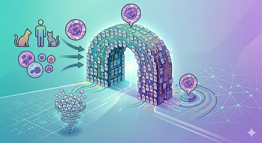
Every now and then we like to share some views “behind the scenes” of our research group, covering topics that haven’t seen the light of day yet. This time, we want to share about the progress of our DFG/FWF-funded project (mu)ROMI – Robust and Accurate Multi-Tumor, Multi-Species, Multi-Laboratory and Multi-Scanner Mitosis Detection with Large-Scale Datasets and Artificial Intelligence.
Driven by the insight that truly robust pattern recognition is mostly always achieved only by having data of sufficient size, quality and (most importantly) diversity, we set out to create the biggest and most diverse mitotic figure dataset, covering many tumor types and loads of cases from human and veterinary pathology.
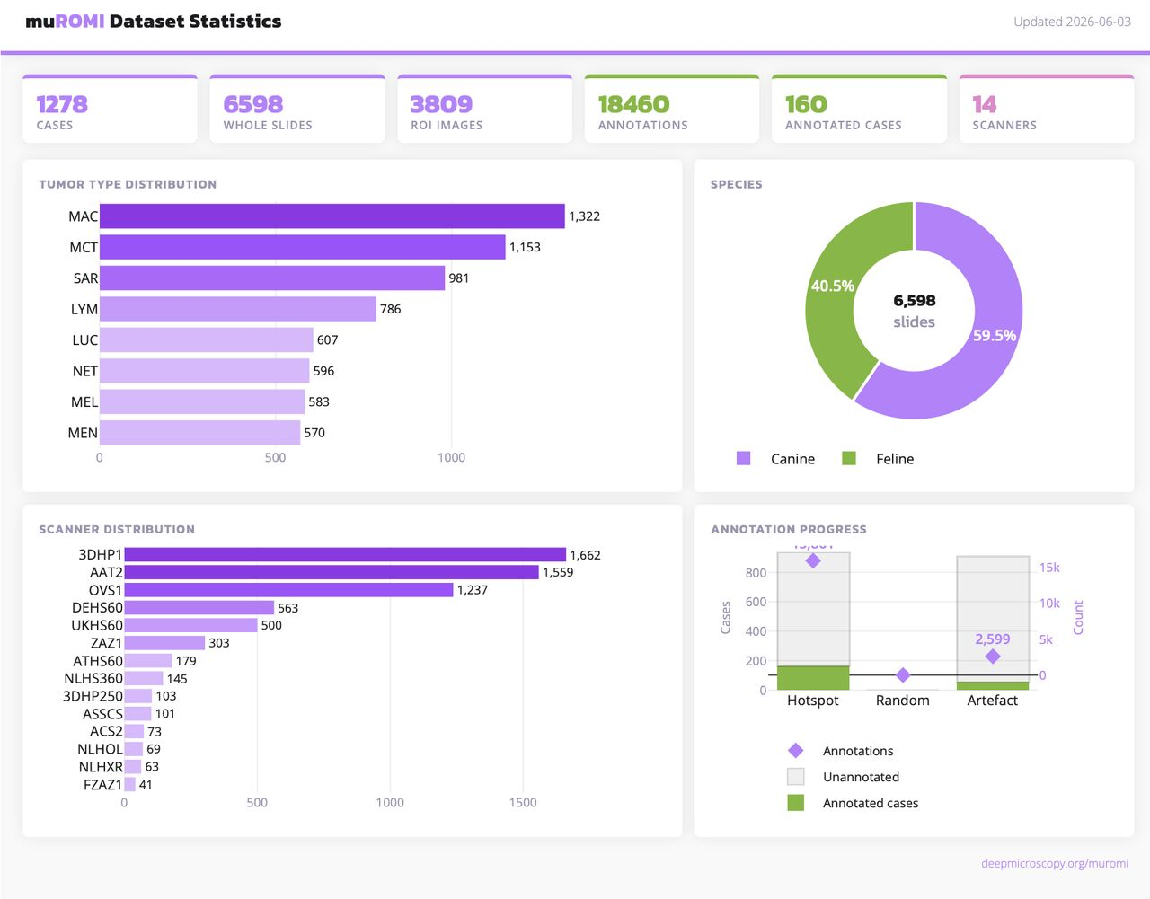
Right now, we have all samples from the veterinary side completed, and are in the midst of the annotation phase. In terms of data, this means that we have digitized around 19 TB of images (mostly WSIs), scanned by various scanners to attribute for scanner domain shift. For each case, we did stain the same very tissue twice (with scanning in-between), using the normal H&E dye and PHH3 IHC stain to highlight mitotic figures.
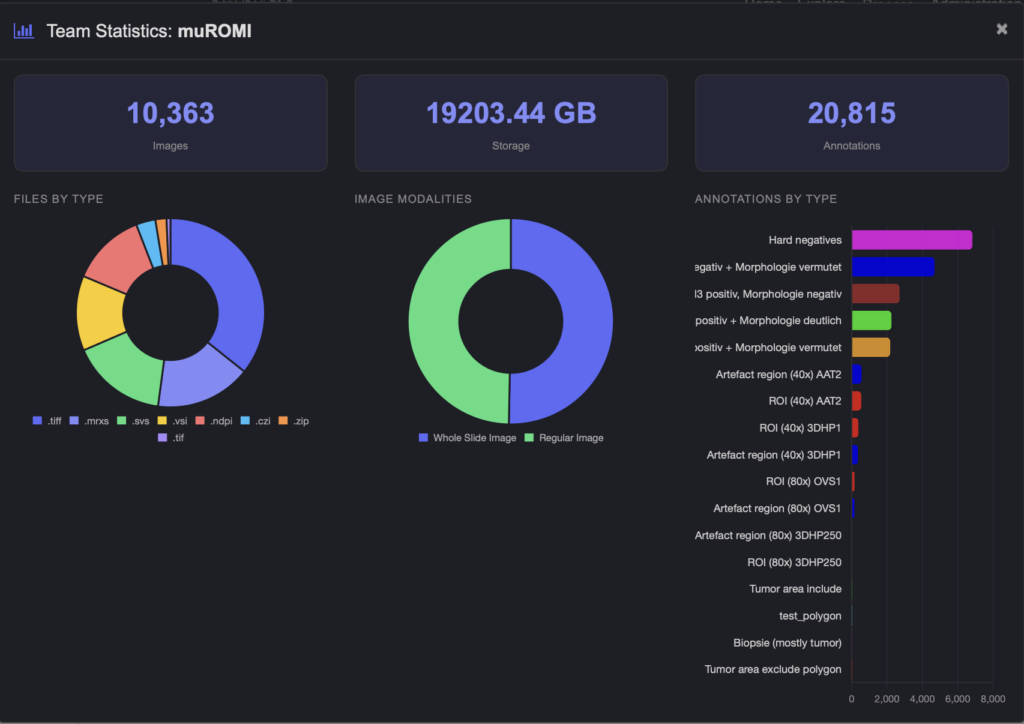
The targets you write into a grant proposal are always challenging to achieve – but at the moment it looks very promising that we will be able to! This is only possible thanks to a great team.
Details about the project here: https://deepmicroscopy.org/muromi

Comments are closed