FOKUS-TML: Optimization of focal planes in tumor diagnostics using multi-z-stack methods
This project aims to enhance the diagnosis of tissue samples, particularly in determining tumor malignancy, through the use of artificial intelligence (AI) methods on digitized tissue sections. While AI has demonstrated improved diagnostic reproducibility, speed, and accuracy, challenges arise in cases with borderline malignancy due to multiple relevant focus planes in tissue sections. Unlike traditional microscopy, digital microscopy captures only one focus setting, leading to the loss of diagnostic information. This issue is prominent in histopathology and further exacerbated in the examination of detached tissue cells (cytopathology). To address these limitations, the project explores three key areas. First, in collaboration with ZEISS Research Microscopy Solutions in Munich, the project aims to robustly detect focus offsets in two-dimensional images, triggering partial re-digitization when the offset exceeds specified tolerances. The second part, in partnership with Tribun Health in Paris, focuses on predicting all diagnostically relevant focus planes from two-dimensional images and recognizing pathological patterns in multi z-stacks. These efforts aim to reduce economic factors and facilitate diagnostic applications in the medical field.
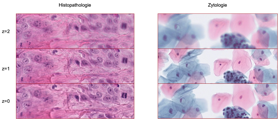
Figure. 1: Pathological specimen contains different valid focus layers.
Publications
In Journals
Emely Rosbach, Jonas Ammeling, Jonathan Ganz, Christof A. Bertram, Thomas Conrad, Andreas Riener, Marc Aubreville: Stuck on Suggestions: Automation Bias, the Anchoring Effect, and the Factors That Shape Them in Computational Pathology. In: Machine Learning for Biomedical Imaging, vol. 3, no. MELBA–BVM 2025 Special Issue, pp. 126–147, 2026.
Sweta Banerjee, Viktoria Weiss, Taryn A. Donovan, Rutger H. J. Fick, Thomas Conrad, Jonas Ammeling, Nils Porsche, Robert Klopfleisch, Christopher Kaltenecker, Katharina Breininger, Marc Aubreville, Christof A Bertram: Benchmarking Deep Learning and Vision Foundation Models for Atypical vs. Normal Mitosis Classification with Cross-Dataset Evaluation. In: Machine Learning for Biomedical Imaging, no. MELBA–BVM 2025 Special Issue, pp. 115–125, 2026.
Jonas Ammeling, Jonathan Ganz, Emely Rosbach, Ludwig Lausser, Christof A. Bertram, Katharina Breininger, Marc Aubreville: Benchmarking Foundation Models for Mitotic Figure Classification. In: Machine Learning for Biomedical Imaging, vol. 3, no. MELBA–BVM 2025 Special Issue, pp. 38–55, 2026, ISSN: 2766-905X.
Sweta Banerjee, Christof A. Bertram, Viktoria Weiss, Jonas Ammeling, Thomas Conrad, Nils Porsche, Robert Klopfleisch, Christoph Stroblberger, Christopher Kaltenecker, Katharina Breininger, Marc Aubreville: Reporting transparency in veterinary pathology deep learning: A systematic review of reproducibility-critical details. In: Veterinary Pathology, pp. 03009858261459452, 2026, ISSN: 0300-9858, 1544-2217.
Jonas Ammeling, Jonathan Ganz, Frauke Wilm, Katharina Breininger, Marc Aubreville: Investigation of Class Separability within Object Detection Models in Histopathology. In: IEEE Transactions on Medical Imaging, vol. 44, no. 8, pp. 3162–3174, 2025, ISSN: 0278-0062, 1558-254X.
Jonas Ammeling, Marc Aubreville, Alexis Fritz, Angelika Kießig, Sebastian Krügel, Matthias Uhl: An interdisciplinary perspective on AI-supported decision making in medicine. In: Technology in Society, vol. 81, pp. 102791, 2025, ISSN: 0160791X.
Jonathan Ganz, Christian Marzahl, Jonas Ammeling, Emely Rosbach, Barbara Richter, Chloé Puget, Daniela Denk, Elena A. Demeter, Flaviu A. Tăbăran, Gabriel Wasinger, Karoline Lipnik, Marco Tecilla, Matthew J. Valentine, Michael J. Dark, Niklas Abele, Pompei Bolfa, Ramona Erber, Robert Klopfleisch, Sophie Merz, Taryn A. Donovan, Samir Jabari, Christof A. Bertram, Katharina Breininger, Marc Aubreville: Information mismatch in PHH3-assisted mitosis annotation leads to interpretation shifts in H&E slide analysis. In: Scientific Reports, vol. 14, no. 1, pp. 26273, 2024, ISSN: 2045-2322.
Marc Aubreville, Jonathan Ganz, Jonas Ammeling, Emely Rosbach, Thomas Gehrke, Agmal Scherzad, Stephan Hackenberg, Miguel Goncalves: Prediction of tumor board procedural recommendations using large language models. In: European Archives of Oto-Rhino-Laryngology, 2024, ISSN: 1434-4726.
Jonathan Ganz, Jonas Ammeling, Samir Jabari, Katharina Breininger, Marc Aubreville: Re-identification from histopathology images. In: Medical Image Analysis, pp. 103335, 2024, ISSN: 13618415.
Marc Aubreville, Nikolas Stathonikos, Taryn A. Donovan, Robert Klopfleisch, Jonas Ammeling, Jonathan Ganz, Frauke Wilm, Mitko Veta, Samir Jabari, Markus Eckstein, Jonas Annuscheit, Christian Krumnow, Engin Bozaba, Sercan Çayır, Hongyan Gu, Xiang ‘Anthony’ Chen, Mostafa Jahanifar, Adam Shephard, Satoshi Kondo, Satoshi Kasai, Sujatha Kotte, V. G. Saipradeep, Maxime W. Lafarge, Viktor H. Koelzer, Ziyue Wang, Yongbing Zhang, Sen Yang, Xiyue Wang, Katharina Breininger, Christof A. Bertram: Domain generalization across tumor types, laboratories, and species — Insights from the 2022 edition of the Mitosis Domain Generalization Challenge. In: Medical Image Analysis, vol. 94, pp. 103155, 2024, ISSN: 13618415.
Conference proceedings
Jonas Ammeling, Jonathan Ganz, Frauke Wilm, Katharina Breininger, Marc Aubreville: Abstract: Investigation of Class Separability within Object Detection Models in Histopathology. In: Handels, Heinz; Breininger, Katharina; Deserno, Thomas; Maier, Andreas; Maier-Hein, Klaus; Palm, Christoph; Tolxdorff, Thomas (Ed.): Bildverarbeitung für die Medizin 2026, pp. 18–18, Springer Fachmedien Wiesbaden, Wiesbaden, 2026, ISBN: 978-3-658-51099-2 978-3-658-51100-5, (Series Title: Informatik aktuell).
Sweta Banerjee, Timo Gosch, Sara Hester, Viktoria Weiss, Thomas Conrad, Taryn A. Donovan, Nils Porsche, Jonas Ammeling, Christoph Stroblberger, Robert Klopfleisch, Christopher Kaltenecker, Christof A. Bertram, Katharina Breininger, Marc Aubreville: Enabling Fast and Mobile Histopathology Image Annotation through Swipeable Interfaces SWAN. In: Handels, Heinz; Breininger, Katharina; Deserno, Thomas; Maier, Andreas; Maier-Hein, Klaus; Palm, Christoph; Tolxdorff, Thomas (Ed.): Bildverarbeitung für die Medizin 2026, pp. 203–209, Springer Fachmedien Wiesbaden, Wiesbaden, 2026, ISBN: 978-3-658-51099-2 978-3-658-51100-5, (Series Title: Informatik aktuell).
Emely Rosbach, Jonas Ammeling, Sebastian Krügel, Angelika Kießig, Alexis Fritz, Jonathan Ganz, Chloé Puget, Taryn Donovan, Andrea Klang, Maximilian C. Köller, Pompei Bolfa, Marco Tecilla, Daniela Denk, Matti Kiupel, Georgios Paraschou, Mun Keong Kok, Alexander F. H. Haake, Ronald R. De Krijger, Andreas F. -P. Sonnen, Tanit Kasantikul, Gerry M. Dorrestein, Rebecca C. Smedley, Nikolas Stathonikos, Matthias Uhl, Christof A. Bertram, Andreas Riener, Marc Aubreville: “When Two Wrongs Don’t Make a Right” – Examining Confirmation Bias and the Role of Time Pressure During Human-AI Collaboration in Computational Pathology. In: Proceedings of the 2025 CHI Conference on Human Factors in Computing Systems, pp. 1–18, ACM, Yokohama Japan, 2025, ISBN: 9798400713941.
Jonas Ammeling, Marc Aubreville, Sweta Banerjee, Christof A. Bertram, Katharina Breininger, Dominik Hirling, Peter Horvath, Nikolas Stathonikos, Mitko Veta: Mitosis Domain Generalization Challenge 2025. In: Zenodo, 2025.
Emely Rosbach, Jonathan Ganz, Jonas Ammeling, Andreas Riener, Marc Aubreville: Automation Bias in AI-assisted Medical Decision-making under Time Pressure in Computational Pathology. In: Palm, Christoph; Breininger, Katharina; Deserno, Thomas; Handels, Heinz; Maier, Andreas; Maier-Hein, Klaus H.; Tolxdorff, Thomas M. (Ed.): Bildverarbeitung für die Medizin 2025, pp. 129–134, Springer Fachmedien Wiesbaden, Wiesbaden, 2025, ISBN: 978-3-658-47421-8 978-3-658-47422-5.
Sweta Banerjee, Viktoria Weiss, Thomas Conrad, Taryn A. Donovan, Jonas Ammeling, Rutger H. J. Fick, Jonas Utz, Robert Klopfleisch, Christopher Kaltenecker, Christof Bertram, Katharina Breininger, Marc Aubreville: Chromosome Mask-Conditioned Generative Inpainting for Atypical Mitosis Classification. In: MICCAI Workshop on Computational Pathology with Multimodal Data (COMPAYL), pp. 266–277, PMLR, 2025.
Sweta Banerjee, Christof A. Bertram, Jonas Ammeling, Viktoria Weiss, Thomas Conrad, Robert Klopfleisch, Christopher Kaltenecker, Katharina Breininger, Marc Aubreville: Comprehensive Dataset of Coarse Tumor Annotations for The Cancer Genome Atlas Breast Invasive Carcinoma. In: Palm, Christoph; Breininger, Katharina; Deserno, Thomas; Handels, Heinz; Maier, Andreas; Maier-Hein, Klaus H.; Tolxdorff, Thomas M. (Ed.): Bildverarbeitung für die Medizin 2025, pp. 260–265, Springer Fachmedien Wiesbaden, Wiesbaden, 2025, ISBN: 978-3-658-47421-8 978-3-658-47422-5.
Jingna Qiu, Nishanth Jain, Jonas Ammeling, Marc Aubreville, Katharina Breininger: Effortless Vision-Language Model Specialization in Histopathology without Annotation. In: MICCAI Workshop on Computational Pathology with Multimodal Data (COMPAYL), 2025.
Christof A. Bertram, Viktoria Weiss, Taryn A. Donovan, Sweta Banerjee, Thomas Conrad, Jonas Ammeling, Robert Klopfleisch, Christopher Kaltenecker, Marc Aubreville: Histologic Dataset of Normal and Atypical Mitotic Figures on Human Breast Cancer (AMi-Br). In: Palm, Christoph; Breininger, Katharina; Deserno, Thomas; Handels, Heinz; Maier, Andreas; Maier-Hein, Klaus H.; Tolxdorff, Thomas M. (Ed.): Bildverarbeitung für die Medizin 2025, pp. 113–118, Springer Fachmedien Wiesbaden, Wiesbaden, 2025, ISBN: 978-3-658-47421-8 978-3-658-47422-5.
Jonathan Ganz, Jonas Ammeling, Emely Rosbach, Ludwig Lausser, Christof A. Bertram, Katharina Breininger, Marc Aubreville: Is Self-supervision Enough?: Benchmarking Foundation Models Against End-to-end Training for Mitotic Figure Classification. In: Palm, Christoph; Breininger, Katharina; Deserno, Thomas; Handels, Heinz; Maier, Andreas; Maier-Hein, Klaus H.; Tolxdorff, Thomas M. (Ed.): Bildverarbeitung für die Medizin 2025, pp. 63–68, Springer Fachmedien Wiesbaden, Wiesbaden, 2025, ISBN: 978-3-658-47421-8 978-3-658-47422-5.
Jonathan Ganz, Chloé Puget, Jonas Ammeling, Eda Parlak, Matti Kiupel, Christof A. Bertram, Katharina Breininger, Robert Klopfleisch, Marc Aubreville: Assessment of Scanner Domain Shifts in Deep Multiple Instance Learning. In: Maier, Andreas; Deserno, Thomas M.; Handels, Heinz; Maier-Hein, Klaus; Palm, Christoph; Tolxdorff, Thomas (Ed.): Bildverarbeitung für die Medizin 2024, pp. 137–142, Springer Fachmedien Wiesbaden, Wiesbaden, 2024, ISBN: 978-3-658-44036-7 978-3-658-44037-4.
Jonas Ammeling, Moritz Hecker, Jonathan Ganz, Taryn A. Donovan, Robert Klopfleisch, Christof A. Bertram, Katharina Breininger, Marc Aubreville: Automated Mitotic Index Calculation via Deep Learning and Immunohistochemistry. In: Maier, Andreas; Deserno, Thomas M.; Handels, Heinz; Maier-Hein, Klaus; Palm, Christoph; Tolxdorff, Thomas (Ed.): Bildverarbeitung für die Medizin 2024, pp. 123–128, Springer Fachmedien Wiesbaden, Wiesbaden, 2024, ISBN: 978-3-658-44036-7 978-3-658-44037-4.
Cookie-Zustimmung verwalten
Um dir ein optimales Erlebnis zu bieten, verwenden wir Technologien wie Cookies, um Geräteinformationen zu speichern und/oder darauf zuzugreifen. Wenn du diesen Technologien zustimmst, können wir Daten wie das Surfverhalten oder eindeutige IDs auf dieser Website verarbeiten. Wenn du deine Zustimmung nicht erteilst oder zurückziehst, können bestimmte Merkmale und Funktionen beeinträchtigt werden.
Funktional
Always active
Die technische Speicherung oder der Zugang ist unbedingt erforderlich für den rechtmäßigen Zweck, die Nutzung eines bestimmten Dienstes zu ermöglichen, der vom Teilnehmer oder Nutzer ausdrücklich gewünscht wird, oder für den alleinigen Zweck, die Übertragung einer Nachricht über ein elektronisches Kommunikationsnetz durchzuführen.
Vorlieben
Die technische Speicherung oder der Zugriff ist für den rechtmäßigen Zweck der Speicherung von Präferenzen erforderlich, die nicht vom Abonnenten oder Benutzer angefordert wurden.
Statistiken
Die technische Speicherung oder der Zugriff, der ausschließlich zu statistischen Zwecken erfolgt.Die technische Speicherung oder der Zugriff, der ausschließlich zu anonymen statistischen Zwecken verwendet wird. Ohne eine Vorladung, die freiwillige Zustimmung deines Internetdienstanbieters oder zusätzliche Aufzeichnungen von Dritten können die zu diesem Zweck gespeicherten oder abgerufenen Informationen allein in der Regel nicht dazu verwendet werden, dich zu identifizieren.
Marketing
Die technische Speicherung oder der Zugriff ist erforderlich, um Nutzerprofile zu erstellen, um Werbung zu versenden oder um den Nutzer auf einer Website oder über mehrere Websites hinweg zu ähnlichen Marketingzwecken zu verfolgen.