
Neoplasia, a common cause of death in both animals and humans, requires treatment decisions based on pathological examination of tumor specimens. Histological analysis, particularly mitotic count (MC), plays a crucial role in prognostication. However, our research highlights significant challenges, including heterogeneity in MC distribution throughout histological slides, observer-dependent variability, morphological complexities, and technical limitations in mitotic figure detection. These factors contribute to poor agreement among trained pathologists, with published κ values between 0.08 and 0.77, posing a risk of incorrect prognostication and therapeutic decisions (Bertram et al., 2018; Donovan et al., 2020).
Our research shows that computer-aided image analysis using machine/deep learning techniques excels in histopathologic classification tasks on H&E-stained slides, surpassing human observers with ample data (Bertram et al., 2022; Bertram 2019a). We’ve developed large-scale, high-quality labeled datasets, and our algorithm has proven to enhance pathologists’ performance significantly (Bertram et al., 2022). This advancement suggests a more reliable tumor prognostication using the cost-effective H&E-stained slides (Villani et al., 2016; Tracht, Zhang, and Peker, 2017).
The need for a multi-domain reference whole slide image data set as open data.
Successful implementation of deep learning-based, data-driven methods hinges on access to representative large-scale datasets. Image domains in histopathology, encompassing staining variations, species, tissue types, and digitization devices, contribute to dataset variability. Different regions within histological sections may contain diverse tissue components and artifacts, potentially rendering selected regions of interest (ROI) non-representative (Aubreville et al., 2020b; Bertram et al., 2019a). This variation leads to a domain shift in data distribution, impacting algorithm performance significantly (Aubreville et al., 2021b; Stacke et al., 2021). Addressing this challenge through domain adaptation approaches is crucial in computational histopathology, and while progress has been made, a persistent domain gap impedes the development of robust software solutions applicable to the full spectrum of biological and imaging variations in digitized histological tumor sections across different tissue types, image locations, laboratories, species, and scanners (Aubreville et al., 2020a; Stacke et al., 2021).
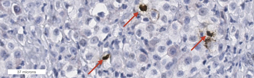
Co-registered H&E and PHH3 stains. The left image shows mitotic figures in an early phase (PHHE-positive) and the right panels show a mitotic figure in a late phase (PHHE-negative).
Dataset Dashboard
We transparently report on our dataset creation progress. Have a look at our dashboard. The full version is available at this link.
Publications
In Journals
- Benchmarking Deep Learning and Vision Foundation Models for Atypical vs. Normal Mitosis Classification with Cross-Dataset Evaluation. In: Machine Learning for Biomedical Imaging, no. MELBA–BVM 2025 Special Issue, pp. 115–125, 2026.
- Benchmarking Foundation Models for Mitotic Figure Classification. In: Machine Learning for Biomedical Imaging, vol. 3, no. MELBA–BVM 2025 Special Issue, pp. 38–55, 2026, ISSN: 2766-905X.
- A Subphase-Labeled Mitotic Dataset for AI-powered Cell Division Analysis. In: Scientific Data, 2026, ISSN: 2052-4463.
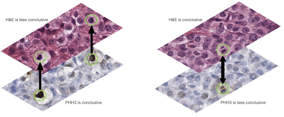
- Information mismatch in PHH3-assisted mitosis annotation leads to interpretation shifts in H&E slide analysis. In: Scientific Reports, vol. 14, no. 1, pp. 26273, 2024, ISSN: 2045-2322.
- Domain generalization across tumor types, laboratories, and species — Insights from the 2022 edition of the Mitosis Domain Generalization Challenge. In: Medical Image Analysis, vol. 94, pp. 103155, 2024, ISSN: 13618415.
Conference proceedings
- Chromosome Mask-Conditioned Generative Inpainting for Atypical Mitosis Classification. In: MICCAI Workshop on Computational Pathology with Multimodal Data (COMPAYL), pp. 266–277, PMLR, 2025.
- Enabling Fast and Mobile Histopathology Image Annotation through Swipeable Interfaces SWAN. In: Handels, Heinz; Breininger, Katharina; Deserno, Thomas; Maier, Andreas; Maier-Hein, Klaus; Palm, Christoph; Tolxdorff, Thomas (Ed.): Bildverarbeitung für die Medizin 2026, pp. 203–209, Springer Fachmedien Wiesbaden, Wiesbaden, 2026, ISBN: 978-3-658-51099-2 978-3-658-51100-5, (Series Title: Informatik aktuell).
- Mitosis Domain Generalization Challenge 2025. In: Zenodo, 2025.
- Comprehensive Dataset of Coarse Tumor Annotations for The Cancer Genome Atlas Breast Invasive Carcinoma. In: Palm, Christoph; Breininger, Katharina; Deserno, Thomas; Handels, Heinz; Maier, Andreas; Maier-Hein, Klaus H.; Tolxdorff, Thomas M. (Ed.): Bildverarbeitung für die Medizin 2025, pp. 260–265, Springer Fachmedien Wiesbaden, Wiesbaden, 2025, ISBN: 978-3-658-47421-8 978-3-658-47422-5.
- Histologic Dataset of Normal and Atypical Mitotic Figures on Human Breast Cancer (AMi-Br). In: Palm, Christoph; Breininger, Katharina; Deserno, Thomas; Handels, Heinz; Maier, Andreas; Maier-Hein, Klaus H.; Tolxdorff, Thomas M. (Ed.): Bildverarbeitung für die Medizin 2025, pp. 113–118, Springer Fachmedien Wiesbaden, Wiesbaden, 2025, ISBN: 978-3-658-47421-8 978-3-658-47422-5.
- Is Self-supervision Enough?: Benchmarking Foundation Models Against End-to-end Training for Mitotic Figure Classification. In: Palm, Christoph; Breininger, Katharina; Deserno, Thomas; Handels, Heinz; Maier, Andreas; Maier-Hein, Klaus H.; Tolxdorff, Thomas M. (Ed.): Bildverarbeitung für die Medizin 2025, pp. 63–68, Springer Fachmedien Wiesbaden, Wiesbaden, 2025, ISBN: 978-3-658-47421-8 978-3-658-47422-5.
Conference posters
- V. Weiss, et al.: Development of a deep learning algorithm for mitotic figure detection in canine lymphoma cytology whole-slide images, ACVP congress 2025
Conference talks
Press coverage
- Imke Stock: Von Hund zu Mensch: KI erkennt Krebs in der Pathologie. Heise online. https://www.heise.de/hintergrund/Pathologische-Mustererkennung-KI-nutzt-Tierdaten-9789792.html